JONES X-RAY

Go Digital Today!

JONES X-RAY OFFERS THE BEST DIGITAL OPTIONS AVAILABLE!
Call Today • 972-647-0171

Stacks Image 10654

DELWORKS DIGITAL RADIOGRAPHY 3.0:


headerumglogo

DELWORKS EDR

An intuitive and powerful DR retrofit system featuring wireless lightweight detectors that are smart and User-Friendly


DELWORKS is a powerful image acquisition and processing software featuring a user-friendly interface that provides sophisticated and speedy medical imaging. Designed with complex system automation, DELWORKS aims to simplify the examination process by making the difficult decisions for you.

Its advanced anatomical programming and image processing algorithms help optimize technologist productivity. DELWORKS strives to minimize human error and unwanted repeat exams to give technologists high quality images at a lower dose, with every exposure.

Cut Image Acquisition Down To Seconds!

Stacks Image 10857


headerumglogo

DELWORKS

Acquisition and Processing Software
Pre-exposure display of patient and procedure information, X-ray generator exposure factors, status and control functions integrated in a single display screen

Exam-specific image processing algorithms
automatically optimize images based on selected patient anatomy

Enhanced image processing parameters.
APR specific default values and manual adjustment, if desired

Image rotation in 90° steps, horizontal mirroring, automatic and manual image cropping to collimated area

Easy verification and correction of image laterality and patient orientation

Intuitively add orientation markers and text comments directly to acquired images (pre-defined or free text)

Detailed histograms of pixel density User selection of modified LUT (Look-Up Table) based on various exam types

Manual adjustment of the LUT, window and level

Effectively manage rejected images
Stacks Image 10678
Image Stitching

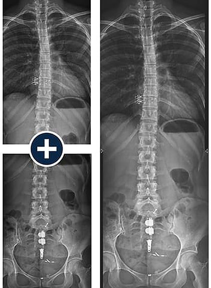
The optional image stitching application
requires no unnecessary preprogramming
or additional equipment, making full-length spine studies effortless and accurate.


Premium Options Available in Single, Dual, or Multi-Detector Applications

Stacks Image 10840
E14C - WIRELESS DETECTOR

The E14C is an ultra-light, portable 14” x 17” (35 cm x 43 cm) wireless detector with outstanding image quality, offering the compact versatility needed to optimize workflow. The E14C has an internal accelerometer which automatically sense motion and takes the detector in and out of ready mode, extending its battery life.

E24C - WIRELESS DETECTOR

The E24C is an ultra-light, portable 24 cm x 30 cm wireless detector with outstanding image quality, offering the compact versatility needed to optimize workflow. The E24C has an internal accelerometer which automatically senses motion and takes the detector in and out of ready mode, extending
its battery life. Due to the compact size, weight, and portability of the E24C detector it is ideal for small anatomy and pediatric imaging.

pdf_sm E14Cw Brochure

pdf_sm E24C Brochure

Stacks Image 10843
E14Ce - WIRELESS DETECTOR

An economical and technology rich 14" x 17" option with Cesium Iodide

The E14Ce is an economical, portable 14” x 17” (35 cm x 43 cm) wireless detector with outstanding image quality, offering the compact versatility needed to optimize workflow. The E14Ce has an internal accelerometer which automatically senses motion and takes the detector in and out of ready mode, extending
its battery life.


pdf_sm E14Ce Brochure


Stacks Image 10846
E17C FIXED DETECTOR

Large format 17" x 17" detector designed to minimize technologist interaction with upright exams in a dual or multi-detector configuration.

The E17C fixed, 17” x 17” (43 cm x 43 cm), large format flat-panel detector is designed to minimize technologist interaction with upright exams in a dual or multi-detector configuration. It is easy to integrate in all types of X-ray systems and delivers both quality images and fast exams, for increased productivity.


pdf_sm E17C Fixed Brochure


Stacks Image 10849
DELWORKS LLI

Monolithic long length 17" x 42" detector designed for full spine and leg imaging in one exposure.

Improve workflow and decrease patient dose with DELWORKS LLI. The extensive image area of 17” x 42” (43 cm x 108 cm) enables full spine and long leg imaging with just one exposure. DELWORKS LLI eliminates potential stitching misalignments for improved confidence in diagnosis. Portability allows upright or supine image acquisition. Developed with highly sensitive AED (Automatic Exposure Detection) technology, DELWORKS LLI can be easily connected and synchronized with any x-ray generator.


pdf_sm LLI Long Length Brochure

EasyConnect​

DELWORKS E-Series Wireless Detectors feature EasyConnect — an Auto Exposure Detection (AED) technology that keeps the detector in a standby mode, awaiting exposure from any X-ray source. Once an exposure is detected it instantly capatures the X-ray image and transmits it wirelessly to the system workstation

Stacks Image 10723
headerumglogo

page0_3-3DELWORKS FIT Anytime, Anywhere

The DELWORKS FIT Portable Tablet Workstation aims to maximize the portability and efficiency of the DELWORKS software — utilizing it anytime, anywhere.

All of the excellent user-friendly features of the DELWORKS desktop software are beautifully translated to an able-bodied tablet PC, capable of withstanding even the harshest radiology (or non radiology) environments. This handheld workstation features wireless connectivity, plenty of storage space, and a powerful Intel® Core™ i5 processor to perform rapid and productive patient side studies, without limitations.

Exis ting analog mobile and portable x-ray devices, radiographic rooms, and other remote or distant locations can greatly benefit from the enhanced adaptability provided by the DELWORKS FIT

iconpdf DELWORKS Fit Brochure


DELWORKS Workstation:
  • Operating system: Windows 10 64-bit
  • Processor: 3.0GHz, Intel Core i5
  • Storage Memory: 16GB RAM, 1 TB HD
Stacks Image 10589

MOVING FROM CR TO DR

  • THE PRACTICAL PERSPECTIVE - 11/21

    MOVING FROM CR TO DR - THE NEXT DIGITAL STEP…

    crtodr-sm

    Before deciding to make the switch from CR to DR, it is important to understand what the differences between the technologies mean to you and your practice. Rather than taking pages and pages answering this question from a technical approach, let’s look at what it means practically.

  • READ MORE

    MOVING FROM CR TO DR - THE NEXT DIGITAL STEP…

    Before deciding to make the switch from CR to DR, it is important to understand what the differences between the technologies mean to you and your practice. Rather than taking pages and pages answering this question from a technical approach, let’s look at what it means practically.

    CR-DR-BIG

    The most valuable resource
    Let’s explore how DR can save your department one of its most precious assets: Time.

    If you currently are working with CR in your department, you have probably worn out a few pairs of shoes with the amount of steps you’ve taken between the bucky and the processor; and you’ve probably become better at shuffling cassettes than a blackjack dealer shuffling cards.

    This is where the first advantage of DR comes in. Rather than swapping cassettes between the bucky and the processor, the image transfers electronically and automatically to the workstation once the x-ray is taken. That’s right, you can just leave the cassette right where it is until the exam is done or you need to move the cassette for another view.

    Flexibility and portability
    The next advantage brings us to your portable x-ray unit. Traditionally, with both film and CR, you didn’t know what your x-ray looked like until you got back to your department. With DR, because your workstation goes with you, there is immediate knowledge on your positioning and technique.

    This is also helpful for those bedside interpretations by physicians, say, in the ICU when they are checking for placement of a line or intubation, the image is visible in seconds, as opposed to waiting for the film to be processed and sent to your PACS for them to see.

    Additionally, if a positioning change is needed, a small adjustment can be made and the image may be repeated, rather than the long walk of shame back to the patient’s room where the entire exam must begin again.

    Lowering radiation dose and achieving higher image quality
    Outside of the time savings to the department and the staff, there is also an arguably more important savings happening with DR when compared to CR, and that is radiation dose.

    Two mainstream DR panel technologies exist today, which are CsI (Cesium Iodide) and Gadox (Gadolinium Oxysulfide). Between the two, CsI requires less radiation to get an equivalent image when compared with Gadox. Depending on which type of DR panel, dose reduction can be as much as 2 to 3 times when compared to CR. Not only are CsI detectors capable of lower dose requirements, they also have the potential for the highest image quality, which will make reads faster and more accurate.

    While the CsI detector is a major part of this equation, the software of the vendor you choose has great influence on the final image, as well.

    The investment mindset
    So, is there any reason not to switch to DR? That comes down to really one thing: upfront cost.

    While cost is always a concern, it is important to consider upgrading to DR as an investment. As mentioned earlier, saved time can directly translate into money saved, usually due to the ability to perform a greater number of exams each day.

    There is also the cost of maintaining CR cassettes to consider. Because the processor disassembles and reassembles CR cassettes with each use, this results in inevitable--and not insignificant--wear and tear. Remember, the DR panel requires no manual processing.

    Avoid reimbursement reduction
    Finally, there is the upcoming Medicare reimbursement reduction slated to begin in this year for facilities that have not made the switch to DR.

    As part of an initiative to encourage U.S. healthcare providers to adopt DR, Medicare began reducing payments for analog x-ray exams in 2017. This year, even sites using CR will begin to see reimbursement fall—first by seven percent for five years, and then by ten percent.

    Changing from CR to DR is a decision every radiology department must consider, eventually, if they haven’t already. At a certain level, the question isn’t as much if, but when, and understanding the differences between CR and DR and even amongst differing DR systems is vital.

Stacks Image 10496
Did You Know… Digital Radiography is a form of x-ray imaging, where digital X-Ray sensors are used instead of traditional photographic film. Advantages include time efficiency through bypassing chemical processing and the ability to digitally transfer and enhance images. Also less radiation can be used to produce an image of similar contrast to conventional radiography. Digital Radiography (DR) or (DX) is essentially film-less X-ray image capture. In place of X-ray film, a digital image capture device is used to record the X-ray image and make it available as a digital file that can be presented for interpretation and saved as part of the patient’s medical record. The advantages of DR over film include immediate image preview and availability, a wider dynamic range which makes it more forgiving for over- and under-exposure as well as the ability to apply special image processing techniques that enhance overall display of the image. The largest motivator for healthcare facilities to adopt DR is its potential to reduce costs associated with processing, managing and storing films. Typically there are two variants of digital image capture devices. These devices include Flat Panel detectors (FPDs), and High Density Line Scan Solid State detectors.

We use cookies to help us improve our webpage - Read more
Just like every site on the Internet -
Learn why
*Note - TCR
SMS Message Statement added 3/7/2023